Guinea pigs, which are brought into the house at the request of the kids, delight with their good nature and funny behavior. An animal with huge eyes, in which there is no membrane that protects the membrane from the penetration of microbes, is often tormented by conjunctivitis. Not knowing how to treat the eyes of a guinea pig, the owners lubricate the mucous membrane with gels, buy drops prescribed for people, which worsens the condition of the funny rodent and is fraught with blindness.
Causes of sore eyes in animals
The absence of a nictitating membrane opens the way for fungi and viruses, bacterial infections, and allergens. Guinea pigs kept at home have eye problems:
- in case of metabolic disorders;
- with vitamin deficiency:
- for autoimmune disorders;
- for congenital anomalies.
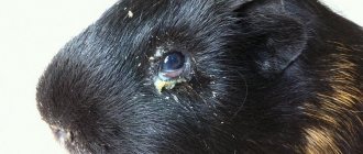
Pus and hyperemia accompany injuries. If the guinea pig is fed improperly, an allergy occurs, which is manifested by squinting. In case of lacrimation, with increasing secretion of white secretion, the animal is taken to the veterinarian, since such signs are present in guinea pigs with eye diseases and pathologies of internal organs. The examination, which is carried out at the Aibolit Plus veterinary clinic, helps to find the cause of pus and lacrimation and prevent loss of vision.
Symptoms
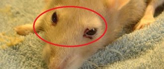
You can miss the onset of the disease only in one case - rarely pick up the guinea pig, approach the cage once every 2 days. In other cases, owners very quickly notice that there is something wrong with the pig’s eyes:
- they become watery and sour;
- clouding of the pupil from the inside or with the formation of a film on the outside;
- redness of the eyelids;
- twitching of the eyeball;
- the eyeball protrudes, turns unnaturally;
- the eyelid turns up and is in an unnatural position.
All these signs are characteristic of different diseases.
| Disease | Causes | Symptoms | Help |
| Keratitis | Injuries, infectious lesions. If the disease occurs in 1 eye, it is likely due to injury. Both are infected | Lacrimation, secretion of mucus, pus. Redness of the eyelid. | Examine the pig's eyes. If there are foreign bodies that have not penetrated the tissue, carefully remove them. If a piece of grass or a pebble penetrates the tissue, immediately contact a veterinarian. Rinse with Furacelin solution, instill drops of Ciprofloxacin, Ciprovet. |
| Conjunctivitis | |||
| Keratoconjunctivitis | |||
| Cataract | Age-related changes, complications after eye injuries, eye infections, as a consequence of diabetes mellitus | The eye crystal becomes cloudy. The pig is losing his sight. | It’s easier to prevent, since cataracts cannot be treated |
| Nystagmus | The result of neurological disorders, most often of the inner ear. Occurs after injuries resulting from a fall, complicated by infections. | The eyeball trembles or moves unnaturally to the side. | Diagnosis by a veterinarian will help determine the root cause, which needs to be treated. |
| Prolapse of the conjunctival sac (“fat eye”) | Occurs as a result of metabolic failure. | The fat fold protrudes above the eyelid and can cover a significant part of the pupil. | It cannot be treated. In rare cases, part of the fat fold is removed with a laser. Such pigs must be excluded from the line of individuals for breeding. |
| Entropione (“turn of the century”) | Genetic disease | The eyelid turns inward. Due to eyelash friction, the mucous membrane of the eye is damaged. | The eyelids are fixed in their normal position with eye ointment. Individuals with entropion are not bred. |
| Copious discharge from the Harderian glands | Physiological norm | A milky liquid leaks from the corner of the eye. This is not pus, but a secret to cleanse the eye. | Excessive secretion is a sign that your pig is experiencing severe body pain. We need to look for its source. The manifestation itself cannot be treated in any way, because it has nothing to do with diseases. |
| Anophthalmia | Developmental anomaly | Guinea pig is born without eyeballs | With proper care, a pig lives and develops in the same way as healthy individuals. |
| Microphthalmia | Reducing eye size | You need to carefully monitor your eyes, because they often accumulate dust. |
Treatment of inflammatory diseases
With conjunctivitis, which develops as a result of injury to the eyeball, when pathogenic bacteria enter the guinea pig's eyelids, the eyelids swell, tears flow, and pus collects in the corners. To relieve inflammation of an animal's eye:
- washed with saline solution;
- instilled with Tsipromed;
- cleared of secretions.
Dried crusts are softened using Albucid and pulled out with tweezers. The skin around the eyes is treated with Tetracycline ointment, which is also placed under the eyelids.
When the cornea is inflamed, the nimble rodent refuses to eat, becomes lethargic, and constantly squints. To cope with keratitis, Solcoseryl ointment is prescribed, which relieves pain and accelerates the healing of microtrauma, and Actovegin is used, which restores the structure of the membrane.
Owners of a guinea pig should begin eye treatment for keratitis immediately; corneal ulceration threatens blindness.
Video
More photos Author(s):
R. Koestlin, prof.
Organization(s):
CTK LMU Muenchen
Magazine:
No. 5-6 - 2013
Translation from German N.A.
Ignatenko EYE DISEASES OF GUINEA PIGS
dermoid
The ocular form of dermoid has been described many times in guinea pigs. Dermoid is most often located on the cornea, especially in the area where the cornea meets the sclera. Recovery without complications occurs after surgical excision of the dermoid and proper comparison of the edges of the eyelids. If the ectopic island of skin irritates the cornea, then lamellar keratectomy is indicated. Additionally, temporary tarsorrhaphy can be performed to protect the eyeball.
anophthalmia (microphthalmos)
Anophthalmia and microphthalmos are severe ophthalmic malformations that have been described in guinea pigs. Intensive anatomical studies of animals with these ophthalmic defects have shown the absence of the optic tract, the absence of Chiasma opticum
(optic chiasm), as well as the absence of the optic nerve. Hypoplasia of the geniculate body of the brain may also additionally be observed. Microphthalmia and corneal dryness, as well as microphthalmia and cataracts in combination with globe rotation, have been described in guinea pigs.
blepharitis
In young guinea pigs, the cause of blepharitis can be a fungal infection (in most cases, trichophytosis caused by Trichophyton mentagrophytes
). In this case, lesions on the head and forelimbs are often observed simultaneously. Local therapy with fungicides such as miconazole, tolnaftate or thiabendazole on paraffin usually leads to successful cure.
eyelid damage
Damage to the eyelids, as a rule, is a consequence of the struggle of animals living together; this problem is most often observed when rabbits are kept together. Less often in the summer, you can observe damage to the eyelids of guinea pigs, which are transferred to enclosures for the summer, and they can injure their eyelids with a metal fence. Surgical treatment of puncture and cut eyelid injuries in guinea pigs follows the same principles as the treatment of eyelid injuries in dogs and cats.
conjunctivitis
Guinea pigs often present to the clinic with conjunctivitis. Various pathogens are isolated from the conjunctival sac: Streptococcus zooepidemicus
,
Staphylococcus aureus
,
Pasteure/la multocida
,
Proteus spp
, however, it is still impossible to say with certainty which of these bacteria are part of the normal microflora of the conjunctiva in guinea pigs and which are pathogenic.
Unilateral conjunctivitis may result from injury, while bilateral involvement is more likely to be part of a systemic disease such as aeromonal septicemia. Especially often, infections affecting the upper respiratory tract, such as Bordetella bronchiseptica
and
Streptococcus pneumoniae,
also lead to eye lesions, in particular conjunctivitis.
A frequently isolated pathogen from the conjunctival sac of guinea pigs is Chlamydia psittaci
. The observed changes include slight hyperemia of the edges of the eyelids to deep purulent lesions of the conjunctival sac. In young animals, this disease is characterized by the formation of inclusion bodies in the epithelial cells of the conjunctiva and is combined with leukocyte infiltrates. In most cases, spontaneous self-healing in sick animals occurs within a month. In addition to infectious causes, conjunctivitis can be associated with violation of living conditions, for example, abundant bacterial contamination of the litter, heavy dust formation, high concentration of ammonia due to insufficient cleaning of cells. A lack of vitamin C also plays an important role in the occurrence of conjunctivitis in guinea pigs. Guinea pigs, like primates, are mammals that require vitamin C from outside, for example, with food. The absence of the enzyme alpha-gulonolactone oxidase makes exogenous supply of vitamin C vital. The first clinical symptoms in guinea pigs appear after about two weeks of lack of vitamin C. The manifestations of this deficiency are varied: it can be weakness, lethargy, diarrhea, anorexia, weight loss and swelling in the joints. A white viscous secretion is observed from the eyes. Therapy consists of eliminating the root cause: replenishing the missing vitamin C (for example, in the form of intramuscular injections of ascorbic acid at a dosage of 50 mg/kg once every three days or daily). Other than eye care (wiping the area around the eyes), no additional eye therapy is required.
protrusion of the orbit/glands (“pea-eve”)
From time to time, a pea-sized subcutaneous (subconjunctival) bulge in the ventral orbit may be seen in guinea pigs. Due to such clinical manifestations, in English this term is referred to as “pea-eve” (or “fatty eye”).
Biopsy studies have shown that these masses may be part of the GI lacrimal glands. Lacrimalis
or
G.I.
Zygomatica . Depending on the size, the protrusion can lead to ectropion (turning out) of the lower eyelid or lagophthalmos (incomplete closure of the eyelids) with subsequent axial degeneration of the cornea. However, as a rule, this condition does not require therapeutic intervention.
corneal opacities
Some guinea pigs may have bilateral oval opacities centered on the cornea. When examined with a slit lamp, gray areas of opacification of varying density were observed in the anterior third of the corneal stroma. Some of these opacities showed some progression, but did not lead to total corneal opacification. Similar-appearing corneal opacities in dogs are described as conditions of corneal degeneration, that is, secondarily developed pathological changes in the cornea. It is assumed that such dystrophic changes in the cornea in guinea pigs are often associated with intraocular ossification. There are still no publications on the cause of such corneal opacities in guinea pigs. It is only known that such corneal opacities in guinea pigs, unlike rabbits and rats, do not lead to corneal calcification.
EYE DISEASES IN DOMESTIC MICE AND RATS
Due to their small size and difficulty in restraining mice and rats, ophthalmic examination of mice and rats requires specific skill. It is best to hold the belly and tail with your palm, and use your thumb and forefinger to fix the animal’s head. A large, open-ended syringe can make it easier to restrain small rodents. The head peeks out from one end of the syringe, and the tail peeks out from the other. It is easy to fix the head with your thumb. Taking frequent breaks from your study helps minimize stress. General anesthesia is necessary in very rare cases.
In mice and rats, the lacrimal system consists of three structures: the intraorbital gland, the extraorbital gland, and the Gardner's gland.
The intraorbital gland is located in the area of attachment of the masseter muscle and is often mistaken for a neoplasm due to its unusual location.
The Gardner's gland is located behind the eyeball and is light pink in color and U-shaped. It has a single exit channel, which opens at the base of the third eyelid. Microscopically, this gland appears foamy due to its higher lipid content compared to other lacrimal glands. This gland is the only mammalian gland that has the merocrine type of lipid secretion. It is believed that the Gardner's gland contains pheromones, which in animals living underground play an important role in caring for the scalp.
In rodents, this gland contains porphyrin, a red-brown pigment, and additionally secretes melatonin, which should function as extraretinal photoreception. Epiphora, or excessive production of tears on the muzzle, often occurs due to eye irritation or obstruction of the nasolacrimal duct and inflammation of the salivary and lacrimal glands. Too much production of these red tears is called chromodacryorrhea and can be confused with blood. Ultraviolet light allows differentiation: porphyrin should glow in ultraviolet light, but blood should not. Eye irritations can occur due to underlying conditions such as keratitis, conjunctivitis, or irritation due to contaminated litter or high levels of ammonia in the environment. Obstruction of the nasolacrimal duct due to malocclusion or overgrowth of the lower chewing teeth. Edema causes obstruction. Dental treatment improves eye condition.
The most common disease of the lacrimal apparatus is dacryodenitis or inflammation of the Gardner's gland. Chromodacryorrhea is the most common clinical symptom in this condition. Dacryodenitis is very often associated with sialodacryodenitis virus (SDAV), which is a coronavirus and is genetically related to murine hepatitis virus, as well as Parker's coronavirus in rats. This virus is a common problem for rats, especially when living in breeder colonies. The virus is highly contagious and spreads very quickly.
Neoplasms of Gardner's gland are quite common in mice and rare in rats. In most cases, these neoplasms are benign, and the most common symptom is exophthalmos.
Conjunctivitis is a common disease in mice and rats. Complicating factors include P. aeruginosa
,
P. pneumotropica
,
Salmonella
,
Streptobacillus moniliformis
,
Corynebacterium kutscheri
,
C. streptococci
,
Mycoplasma pulmonis
, mousepox virus or lymphocytic choriomeningitis virus.
In one study of aged rats with exophthalmos, the most common cause of conjunctivitis was inflammation of the Gardner's glands. Orbital neoplasms, such as poorly circumscribed adenocarcinoma, solid carcinoma, and poorly circumscribed sarcoma, were rare causes.
corneal dystrophy
Dystrophy is the most common corneal change in rodents and results in a white, dense opacification. In one laboratory, 8 to 10 elderly rats suffered from corneal degeneration, which manifested itself as localized small white dotted opacities. This pathology has remained unchanged since the initial changes. In the same laboratory, mice were more likely to develop corneal dystrophy with age. These changes usually had a geometric shape and were located in a thin layer in the anterior part of the corneal stroma. The pathogenesis is not precisely known, but it is assumed that dystrophic changes in the cornea may be a consequence of keratitis.
Keratitis is regularly observed in laboratory rats and is most often seen in two diseases: SDAV and suppurative keratoconjunctivitis, with SDAV being the more common pathology.
anterior chamber of the eye (choroid)
Iris coloboma often appears as a hole in the iris or a defect at the border of the pupil. Many studies conducted on young mice with coloboma have shown that the coloboma is usually located in the ventral quadrate of the iris and results in a small, ventrally located pupil. Synechiae have also been observed in young and adult mice and rats. Synechiae and uveitis are uncommon in rats, although uveitis is a common problem in older rats.
As in other animal species, uveitis can occur as a result of ocular damage and also possibly as a consequence of systemic diseases.
Spontaneous intraocular neoplasias are relatively rare in mice. In rats, the most common neoplasm is melanoma.
lens
Animal experiments have made it possible to understand the physiology of the lens and the pathogenesis of cataract development. Many different variations of cataract mutations have occurred in mice and rats due to radiation or exposure to a chemical mutagen.
posterior chamber of the eye
Mice and rats have a large spherical lens. This, during ophthalmoscopy, provides a picture of the fundus in which the retina appears to be mobile within the vitreous. Due to their very small size, it is very difficult to examine the fundus in mice; this is done under a +30 to +40 D magnifying glass. Photographing the fundus of the eye is very difficult to take in mice due to the very small size of the pupil and the relatively flat surface of the cornea. Compared to mice, the fundus of rats is easier to examine and photograph.
When using a direct ophthalmoscope to evaluate the fundus, good visibility at +8D magnification is required.
Persistence of hyaloid arteries is very common in growing rats and mice and disappears with age. If fragments remain in adulthood, they may float freely, associated with bleeding (which usually resolves completely) or remain hanging on the posterior capsule of the lens. These cataracts appear as small, granular plaques that usually remain unchanged.
Retinal degeneration has been intensively studied in mice and rats. Some breed lines of mice and rats may have hereditary retinal degeneration, while in other breed lines retinal detachment always occurs secondary to other systemic diseases.
Phototoxic retinopathy is the most thoroughly studied and common cause of secondary retinal degeneration in laboratory mice and rats.
Predisposing factors for the development of phototoxic retinopathy are older age, the concentration of certain hormones, the degree of ocular melanosis, nutrition, genetic factors, duration of light intensity and periodic comparison with constantly switched off lights. Albino mice and rats are genetically more sensitive to the effects of light than more pigmented breeds. It would be nice to determine this condition so that it can be used at home.
Rats with high intraocular pressure may develop associated retinal hemorrhages. Rats with high intraocular pressure may also experience pupillary swelling.
Many congenital ocular pathologies have been described in various strains of mice and may possibly also be observed in domestic mice. The most common congenital pathology is microphthalmia. Alone or in combination with cataracts or retinal dysplasia, microphthalmia occurs sporadically in rats. It is believed to be of genetic origin. Microphthalmia can be confused with phthisis (atrophy) of the eyeball; the latter condition can occur due to eye trauma or previous severe uveitis.
glaucoma
Rats generally have lower intraocular pressure when the light is on and higher intraocular pressure when the light is off. Glaucoma, or increased intraocular pressure, is very rare in rats.
Fighting retrobulbar abscess
Sometimes in a rodent a long tooth root extends beyond the boundaries of the canal and extends into the orbit. During ingrowth, the apple increases in size and protrudes outward. After an X-ray of the visual organ confirms a retrobulbar abscess, our doctors grind down or extract the tooth, and in severe cases, remove the affected eye.
When the eyelids turn up, the eyelashes rub against the cornea, and the membrane becomes cloudy. The pathology cannot be cured, but to eliminate eye pain in a guinea pig, cilia are attached to the upper eyelid with an adhesive composition.
Cartilage or bone formations, which make themselves known as a light stripe inside the eye, grow, which ends in the destruction of the nerve responsible for vision, but it is impossible to stop the process.
Discharge from a boy guinea pig
In male piglets, puberty occurs quite early - at the age of 4-8 weeks. They begin to sniff the females' droppings and mark their territory by secreting a sharp-smelling liquid. Also, white contents (remnants of sperm) may be released from the boy’s genital organ. This is normal.
However, it is important not to confuse the symptoms and not to miss inflammation of the genital organ, which occurs due to contamination with smegma or hair.
The most reasonable decision that a guinea pig owner can make is not to guess on coffee grounds, but to call a ratologist at home. Only a specialist can make an accurate diagnosis and prescribe effective treatment. For your convenience, the Hermes veterinary hospital offers the service of a doctor visiting your home at a convenient time.
Take care of your little furry pet, and he will thank you with a cheerful appearance, fun activities, and a long healthy life!
Changes in the lens and cornea
With advanced keratoconjunctivitis, if treatment for an infectious eye disease is ignored, the cornea of the seagull becomes cloudy, which is manifested by the appearance of a cataract, and dangerous loss of vision. The problem is eliminated in the clinic surgically.
When metabolism changes or there is a lack of vitamins, the lens does not refract light and does not direct rays to the retina. When the eyes are damaged by cataracts, the animal begins to see worse. Treatment slows the onset of blindness.
For glaucoma, which occurs when the cells lining the optic nerve die in a pet:
- the cornea swells;
- the eye swells;
- the pupil does not react to light;
- Tormented by lacrimation.
At the beginning of the appearance of deviations, drugs are instilled to help maintain intraocular pressure at a normal level.
When the conjunctival sac protrudes, which passes to the rodent from its parents, plastic surgery is performed using a laser.
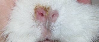
Lichen
Another very unpleasant and dangerous disease, the causative agent of which is a fungus. Ringworm affects the hair follicle and skin.
Signs
The presence of lichen can be determined by several signs:
- in the affected areas, the fur falls off and red, encircling spots appear;
- the skin peels and constantly itches and hurts;
- the appearance of rough, sometimes raised spots on the skin;
- the skin may become scabby;
- the animal is constantly itching.
Kinds
If lichen is not treated, the probability of infection in healthy pigs is almost 100%. There are two types of lichen, which most often affect these particular pets:
- microsporia;
- trichophytosis.
Spores of these fungi can be active for 2-3 years. They are stored in soil, wool and are distributed safely in the environment. Most often, animals with weak immunity and living in unsanitary conditions are susceptible to infection with lichen.
To detect the fungus, a thorough examination is carried out - shining the affected area under a UV lamp and taking skin scrapings in areas of peeling.
Treatment
Sick animals are moved to a separate cage. By completely sterilizing the home and all its accessories. In affected areas, the hair is completely cut off.
To treat lichen, antifungal medications for guinea pigs are used - Chlorhexidine, Clotrimazole, Miconazole. As veterinary practice shows, there are no better drugs today, since the positive effect of treatment is achieved quite quickly.
Types and stages of development of hordeolum
Depending on the location of the lesion, it can be of the following types:
- External stye
. More common according to statistics. The infectious-inflammatory process is localized at the edge of the eyelid, since the infection settles in the sebaceous gland of Zeiss or in the ciliary bulb. - Internal stye on the eye
. It has another name - meibomite. It is caused by the penetration of pathogenic microorganisms into the passage of the meibomian glands located on the back side of the eyelid margin.
By type they distinguish:
- Hot barley
. It is characterized by the classic development of the disease; infection is localized in the ciliary pocket - the bulb or Zeiss gland. With therapy it goes away in about a week. - Cold stye (chalazion, meibomian cyst)
. The inflammatory process affects the meibomian glands. It develops very slowly. Recovery takes 1–2 months. If there is a very large stye, surgical removal is possible.
Inflammation goes through the following stages:
- Formation of a purulent core
. A small reddish swelling causes discomfort when moving the eyelids. - Formation of an abscess
. The patient’s condition at this stage can only be alleviated by medications. - Breakthrough of pus.
It does not indicate recovery, but the patient feels much better. During this period, you need to keep your eyes clean and use prescribed medications and ointments.
Preventive measures
Prevention includes following the recommendations:
- Strengthening immune forces
. This should always be done, not only when there is a threat of hordeolum or in the off-season. It is recommended to take vitamins, give preference to fresh vegetables and fruits, move more, be in the fresh air, and follow a daily routine. - Timely elimination of ophthalmological disorders
. If you are prone to eye diseases, you should regularly visit an ophthalmologist. Self-diagnosis and self-medication usually contribute to the transition of pathology to a chronic form. - Compliance with hygiene standards
. You need to have your own towel, wash your hands with soap, change bed linen every 1–2 weeks, and not use other people’s cosmetics. - Developing the habit of not touching your eyes
. This should not be done even in a normal state, when a person is not bothered by any problems. This rule also applies to the face - you should not touch it during the day with unwashed hands.
Simple preventive measures will help avoid inflammation and keep your eyes healthy.
Is stye contagious?
It is not dangerous for others, but only if basic hygiene rules are followed. The risk of infection usually occurs in young children. Due to their age, they do not yet understand the importance of hygiene and therefore often suffer from bacterial infections. Infection is also possible when the patient uses cosmetics. But in general, isolation of an adult or child suffering from hordeolum is not required.
The causative agents of the disease are found in pus located in the barley sac. There are no bacteria on the surface. But there is usually little pus inside, so even a burst stye is not dangerous. In 80% of cases, the abscess breaks out at night when the patient is sleeping. If purulent masses end up anywhere, it is on a person’s face, his pillow, or bed linen. The infection does not spread further.
Ways to see a pig in the dark
Like other rodents, guinea pigs move well and see in dark rooms. Although they are not actually helpless at night and can perform basic activities, rodent vision is still considered daytime. For this reason, it is ideal to place the cage in a well-lit area, but away from direct sunlight.
Experiments have been conducted to prove the negative effects of darkness on animals. Degradation, disease and various types of mutations occur. It has been observed that in the process of survival, animals navigate better through hearing and smell than through vision.
The most important senses for all rodents are hearing and taste. For this reason, guinea pigs often taste everything, even inedible objects (paper, sawdust, wire, fabric). When searching for food, guinea pigs are guided by the color, smell and taste of food and can remember what they ate (bitter, sweet and salty).
Sweet foods are always preferable. Rodent owners should know that honey and molasses are among the favorite treats of these animals.
The pig can also see in the dark, but much worse than during the day. Rodents are less active at night, but daylight is extremely important for them.
Treatment of eye diseases
Without prior consultation with a veterinarian, no treatment should be carried out, since this can only worsen the situation. Before using corticosteroids, you must make sure that the cornea is not damaged, since such drugs interfere with its recovery and are therefore contraindicated in such situations. The condition of the cornea is checked using special test strips containing fluorescein, which colors the damaged area yellow.
Before treating a sore eye, it is necessary to remove pus and dirt so that the effect and concentration of medications does not decrease. To do this, use warm 0.9% saline solution or acetylcysteine, which dissolves mucus. The discharge can be removed with a cotton pad, which is pre-moistened with a furatsilin solution or ordinary boiled water. Please note that tea leaves and chamomile decoction are strictly contraindicated, as they dry out the mucous membranes and promote the development of fungi and bacteria.
Eye diseases of guinea pigs, the symptoms and treatment of which are closely interrelated, require special attention from a veterinary ophthalmologist. An important step is the restoration of the cornea with the help of vitamins A and C. Artificial tear preparations are indicated to eliminate dry eye. Antibacterial ointments and drops are used up to 4 times a day, sometimes more often.
Serious eye injuries require the following medications:
- "Tobrex";
- "Tsiprovet";
- "Levomycetin";
- "Tsipromed".
If the cornea is injured, the following medications are added to the above:
- "Korneregel" (gel);
- "Solcoseryl" (gel);
- "Balarpan" (drops).
Possible problems with the organs of vision
Despite the underdeveloped organs of vision, pigs do not have any problems with vision. It is rare and is usually a side effect of respiratory problems. Such problems may also indicate diabetes, dental disease, and general dehydration of the animal.
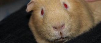
Signs of vision problems include discharge from the eyes and cloudy pupils. The quality of general vision decreases, and the animal is less oriented in space.
Due to deterioration of vision, the animal may begin to develop ulceration of the cornea of the eye. If the eyes are swollen and watery and your pet is acting restless, contact your veterinarian. Cataracts are caused by age-related changes or diabetes.
It happens that small animals are born blind, but thanks to well-developed other organs, they do not differ from their counterparts in their good quality of life and care.
Prevention
Veterinarians recommend following these simple rules:
- choose the right diet for your pet;
- keep the hay clean and the water fresh;
- to sharpen teeth, give your pet hard wood;
- place the cage away from dampness and drafts, keep it clean;
- do not allow the animal to become hypothermic, overheated or wet;
- let your guinea pig run around, this is good for its health;
- control the length of your pet’s claws so that they do not grow too long.