Information / Excessive tooth growth in chinchillas - malocclusion
What are the problems with chinchilla teeth?
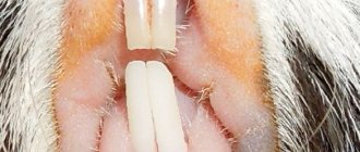
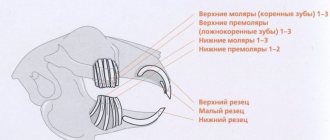
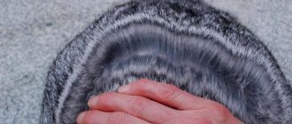
There are twenty teeth in the chinchilla's mouth - 4 front incisors and 16 molars. You can see the front incisors with your own eyes by pulling back the chinchilla’s front and lower lips. The molars are located deeper in the mouth and can only be seen using a special medical device called an otoscope (a funnel with a built-in LED).
Why do chinchilla teeth grow quickly? Because rodents eat dry and solid food. The mechanism of rapid tooth growth is laid down by evolution. Imagine that a chinchilla's teeth grow just like a person's fingernails. A person regularly trims his nails with scissors, otherwise they will grow so long that they will not allow him to work with his hands. A chinchilla cannot “trim” its own teeth; it wears them down by chewing hay, wood, a grindstone and everything else that comes “to the tooth” - a rodent is a rodent.
Teeth regrowth occurs if the chinchilla’s diet does not contain enough roughage and solid food - hay, chewing sticks, whole grains. The teeth grow long, become hooked, and injure the tongue and cheeks. The animal is unable to close its mouth, cannot chew food and dies from hunger. However, do not be discouraged, the problem can be prevented and successfully treated, especially in the early stages.
How many teeth does a chinchilla have, features of their structure
Chinchillas have 20 teeth in total. In the front part of the jaw there are 4 incisors (2 above and 2 below, opposite each other). The front side is characterized by a rich coating of enamel, with a color range from light yellow to orange.
The back side of the incisors is more susceptible to abrasion and is not covered with enamel, which is expressed in the peculiar tip of the incisor, shaped like a chisel. The increase in the size of the teeth at the base is designed to gradually erase the crown of the incisors. If the incisors are not worn down, tooth growth continues and reaches enormous sizes. The length of the tooth crown is 0.6-1.2 cm, the tops of the incisors overlap each other - the upper ones behind the lower ones.
The space between the incisors and molars is called the diastema. The group of cheek teeth is formed by 16 small and large molars:
- small molars (premolar) - 4 pcs.;
- large molars (premolar) - 12 pcs.
The molars are located in proportion to each other, touching occurs over the entire surface, with a total length of about 1.2 cm.
Symptoms of malocclusion
1. The first sign is that the chinchilla becomes “picky” in food. The animal sits at the bowl for a long time, chooses softer food, digs around, and scatters the food. The chinchilla rubs its cheeks with its paws, reduces motor activity, and sometimes sits down for a long time. If you notice changes in eating behavior, start monitoring your pet's weight. Constant weight loss, even small, by 1-2-3 grams per day, is a signal. For an adult chinchilla, losing 10 grams of weight per day is already critical. If you do not observe any digestive problems, then the likely cause is teeth.
2. As the condition develops, both teeth and tooth roots are susceptible to regrowth. Overgrown roots of the upper molars irritate the lacrimal glands. Because of this, the chinchilla's eyes water and his nose also runs. And the sprouted roots of the lower molars put pressure on the salivary glands. The chinchilla is drooling and snot, his face is wet. Therefore, the dysfunction is sometimes called “slobber.”
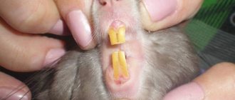
3. In an aggravated condition, you can see how overgrown incisors, curved by hooks, protrude from the chinchilla’s mouth. The animal's mouth does not close because the jaws cannot close. The chinchilla is noticeably losing weight. Teeth and tooth roots, growing into soft tissues, cause inflammation and abscess . Read the article about abscess on the website>>>
The course of the condition is accompanied by progressive weight loss.
Symptoms of dental pathologies in chinchillas
Characteristic symptoms of dental diseases:
- excessive salivation;
- swelling of the jaw;
- copious discharge from the nasal cavity and eyeballs;
- excessive length of incisors;
- partial or complete loss of appetite, consequently, rapid loss of body weight;
- itching in the cheek area;
- "lethargic" behavior;
- diarrhea, diarrhea;
- squealing while eating;
- Frequent brushing of teeth.
At the first signs of illness, it is necessary to urgently contact a specialist in order to avoid further deterioration of the condition and loss of pets.
Pyometra
This terrible disease can be a complication of endometritis and occur independently. When it occurs, pus or other liquid accumulates in the uterine cavity: mucus (mixometra) or water (hydrometra). In the open form, pus flows from the uterus through an open loop as a brown, viscous liquid. If the loop is closed, pus accumulates inside and the uterus swells; the tumor can even be felt in the lower abdomen. Like vaginitis, this disease is contagious, including for males. Fortunately, pyometra and its variations can be diagnosed quite well yourself. Symptoms: refusal to feed and insatiable thirst, as with endometritis, lethargy, apathy, vomiting, frequent and copious urination, swollen abdomen.
All of these diseases can be treated with medication, but serious cases will require surgery and removal of the uterus. In order to prevent the death of an animal, you need to be attentive to it, take care of the quality of food and water, bathe it in the sand and introduce new relatives with great care.
Causes of dental pathologies in chinchillas
Root cause of dental diseases:
- the diet is not balanced;
- lack of solid food, as a result, incomplete abrasion of teeth;
- injuries received that led to the displacement of teeth and the formation of malocclusion;
- genetic diseases;
- congenital pathologies;
- diseases not related to teeth that affect immunity and appetite.
Do you often give your chinchilla solid food?
There are 4 main dental diseases in chinchillas
Ingrown tooth roots
A fairly common pathology is ingrowth or elongation of roots; this problem can develop equally on both jaws. If the ingrowth occurs in the upper jaw, the sinuses and eyes may suffer. If the problem began in the lower jaw, then swelling of the entire jaw is noted.
This disease has a number of characteristic signs:
This pathology is considered very rare and can only be detected using x-rays. This disease has no cure.
Types of dental diseases in chinchillas
The most common dental diseases in rodents are:
- malocclusion;
- crown regrowth;
- ingrown tooth roots;
- tooth loss.
It is not recommended to self-medicate, and at the first signs of dental disease, the help of a qualified specialist is necessary.
Malocclusion
Malocclusion is a disease associated with malocclusion. Excessive size of incisors due to insufficient grinding with the formation of hooks.
Indicative characteristics of this disease are:
- deterioration of appetite and, as a result, weight loss;
- eating mostly soft foods in small pieces;
- tearfulness and vomiting.
As the disease progresses, salivation increases, and the oral cavity is affected by bleeding ulcers. Overgrown teeth injure the rodent's tongue. Only timely specialized assistance will help avoid death.
After cutting teeth, during the rehabilitation period the animal must be helped to eat. Until the pain goes away and the animal can feed on its own, it is necessary to inject the ground food into the oral cavity with a syringe.
To avoid the development of this disease, many experts advise:
- constant monitoring of chinchillas’ nutrition, including granulated food, salt and mineral stones, tree branches;
- Hay is added to the diet, which is rich in fiber and, when chewed, wears down the molars.
How is the bite correction procedure performed?
Often, pet owners worry about how their pets will cope with teeth trimming procedures. I prefer to perform bite correction without the use of anesthesia. The slight stress that an animal may experience during trimming is not commensurate with the stress that your pet experiences with constant pain associated with improper growth of teeth that injure the oral cavity. When correcting the bite, rodents and rabbits do not experience toothache; this manipulation is comparable to trimming their claws, if the doctor has the necessary experience in carrying out the procedure. The procedure itself takes only a few minutes.
Unfortunately, as I said above, it is not always possible to completely eliminate the problem so that bite correction is never required again. If the bite is seriously compromised due to late treatment or improper treatment, further trimming may be required throughout life. Let's imagine that at the age of 2, a guinea pig developed dental problems, and now she will have to trim her teeth 1-2 times a month. Let’s imagine that this pig lives safely for up to 6 years, because it will be taken care of and regularly brought in for bite correction (yes, such animals and such owners really exist!). Over the course of 4 years, you will have to carry out this procedure 50 to 100 times. Regular use of anesthesia for this procedure, even the safest one, can significantly shorten the life of your pet. We are talking about a healthy animal that has no other problems other than dental ones. And if we talk about patients with pathologies of the respiratory system, heart failure, severe liver and kidney diseases, severe exhaustion, anesthesia may be completely contraindicated and lead to death. Therefore, if it is possible to carry out a correction without anesthesia, I always do it. Anesthesia is necessary only in very rare cases.
I perform dental manipulations on chewing teeth using specialized German instruments designed specifically for rodents and rabbits. Unlike Pakistani analogues, non-specialized surgical and manicure instruments, my instruments do not crumble teeth, do not leave cracks in them, do not injure the oral cavity and allow you to achieve the desired result with high precision.
How to trim chinchillas' teeth
It is not recommended to carry out this procedure with improvised means and at home, but you should consult a specialist, since this is a serious operation that is performed in a hospital, using intramuscular injections and anesthesia.
The animal is fixed in a specialized machine to avoid injury to the oral mucosa. The operation is performed by medical personnel using dental fur. grinding, under the guidance of an experienced veterinarian, without the presence of strangers.
ATTENTION! With subsequent regrowth of crowns and the formation of hooks, it is recommended to trim the teeth once every 3-4 months with laser grinding.
To reduce the risk of subsequent possible relapses, you must adhere to some recommendations:
- periodic medical examination by a veterinarian;
- independent control of the oral cavity;
- prompt response: at the first sign of problems, contact the clinic;
- maintaining a proper diet: the presence of solid feed, wood and mineral stones.
During the postoperative period, the pet owner must organize rehabilitation care:
- treat the oral cavity with an antiseptic and herbal decoctions;
- use anesthetics;
- if there is no appetite, feed liquid food from a syringe;
- organize periodic medical supervision.
When health returns to normal, it is advisable to change the diet of your favorite animal. Chinchillas need to consume large amounts of hay and solid food to allow normal tooth wear to occur.
You should visit the veterinarian periodically to prevent possible diseases.
One of the most common problems in herbivorous rodents (chinchillas, guinea pigs, degus) and rabbits that people come to our clinic for is dental disease. These animals' teeth grow throughout their lives. Changes in the shape or position of the teeth lead to malocclusion, that is, a discrepancy between the surfaces of the teeth in the upper and lower jaws. The teeth become curved, sharp protrusions appear on them, which injure the mucous membrane of the mouth. In addition to the growth of the visible crown of the tooth, the reserve crown (root) also grows, which often leads to the formation of abscesses, tooth loss, narrowing or inflammation of the nasolacrimal duct, and osteomyelitis (inflammation) of the skull bones.
Causes
The most common cause of such diseases is hereditary malocclusion. Hereditary diseases most often first appear at 1-2 years of age. Recently, the number of such patients has increased. The age of the youngest patient with dental disease encountered in our practice was 7 months.
In addition to genetic predisposition, there are also acquired causes: improper feeding (lack of coarse fiber in the diet, large amounts of grain and vegetable feed), calcium metabolism disorders, diseases of the temporomandibular joint, head injuries.
Symptoms
Most often, the first thing owners notice is
this is what the animal wants to eat, but cannot - it takes the food and immediately spits it out, or tries to choose softer food components, refusing hay and grass pellets, and twig food.
You may also experience drooling, gradual weight loss, lack of feces or a decrease in size, deterioration in the quality of fur, excessive shedding, discharge of pus from the eyes, swelling (abscesses) on the jaw, disproportion of the muzzle (bulging out of one of the eyes), changes in the occlusion of the incisors (incisors). stop closing correctly, begin to grow, and bend).
Diagnostics
If you experience any of the symptoms listed above, you should schedule an examination with an exotic animal specialist. If recording is not possible immediately, and the animal cannot eat, then it is necessary to start force-feeding the pet from a syringe without a needle with hay crushed in a coffee grinder, mixed with water or herbal granules soaked in warm water. Feeding is carried out every 2-4 hours and should be at least 10 ml of mixture at a time.
At the appointment, the doctor will collect data about the animal, conduct a clinical examination and, if dental disease is suspected, prescribe a dental examination and x-ray examination of the skull.
Dental examination is carried out only under general anesthesia. It is very important that the animal is relaxed. Without induction of anesthesia, when trying to open the mouth with the help of instruments designed for this (mouth and cheek retractors), there is a high risk of fracture of the incisors or dislocation of the lower jaw joint, and the animal twitches, moves the tongue and gnaws on the instrument, which undoubtedly worsens the quality of the examination.
Sometimes the initial examination without anesthesia is performed using an otoscope with a long attachment. This is safe, but also does not allow you to examine the entire oral cavity and reveals only some pathologies.
The problem was easily discovered during the initial examination of this guinea pig or this chinchilla.
Whereas in this rabbit, only an examination under anesthesia helped to make a diagnosis.
At first glance, the teeth look normal, but the second photo shows that upon detailed examination, there is a hook on the far cheek tooth, which has long injured the mucous membrane of the tongue (photo 3 after grinding the hook, the tongue injury is indicated in red).
An X-ray examination is prescribed to assess the condition of the reserve crowns (tooth roots) or to identify the tooth from which the jaw abscess appeared. Such teeth must be removed. An X-ray of the skull is taken in at least 4 projections.
An example of an x-ray of a chinchilla.
In the photo, the first cheek tooth is indicated in red. It can be seen that the condition of the root of this tooth is different from the others, it is being destroyed, there are no clear boundaries. It is also clear that the tooth has deviated from the rest in the row and there is no visible crown. In this case, the remains of the first tooth in the upper jaw were removed and the length of the remaining teeth was corrected. Without the help of x-rays, the pathology would remain undetected, which would subsequently lead to osteomyelitis and, probably, osteolysis (inflammation and destruction of the bones of the skull).
Treatment
Treatment consists of filing down overgrown teeth with a special device. We use a dental micromotor with various bur attachments. Treatment is carried out immediately after a dental examination under general anesthesia. Even if the diagnosis of “dental disease” is made during the initial examination, teeth filing without anesthesia is not carried out, because there is a high risk of injury to the oral mucosa, which in turn can lead to heavy bleeding and death of the animal.
Our clinic uses inhalation anesthesia with the drug “Isoflurane”. This is the safest type of anesthesia for rodents and rabbits. Within 5-15 minutes after the procedure, the animal completely recovers from anesthesia and is ready to go home.
Dental disease is a disease that will recur because... teeth grow constantly, and the correct bite due to changes in the position of the teeth is almost impossible to restore. The intervals between dental corrections are individual and can range from 3 weeks to 1 year.
In addition to filing down overgrown crowns, it may be necessary to remove damaged teeth, open abscesses that have formed, etc.
After correction of dental crowns, rehabilitation therapy is carried out. It may include the prescription of painkillers and anti-inflammatory drugs, long-term antibiotic therapy in the presence of ostemyelitis or purulent processes, correction of intestinal stasis (increasing intestinal motility, combating bloating), diet, force-feeding, parenteral fluid administration (droppers). The prescription of a particular treatment method will depend on the condition of the individual patient.
In our network of clinics, the diagnosis and treatment of this pathology is carried out by an exotic animal specialist, Lyudmila Ivanovna Bazhina. Reception is conducted at the address Perm, st. Turgeneva, 12. Phone for appointment 265-51-99.
Why does a chinchilla grind his teeth?
Grinding of pets' teeth during normal activity and undisturbed appetite is a normal physiological process that indicates wear on the pet's molars. Some individuals do this even while sleeping.
When grinding and chattering of teeth occurs with poor or completely absent appetite, abnormal stool, excessive salivation, deterioration in the functioning of the limbs, emergency assistance from specialists is needed to avoid the premature death of your animal. All this may indicate food poisoning of the animal.
Classification: types of main dental diseases of the teeth
Conventionally, all violations can be divided into two broad categories - those associated with damage to the enamel, and those caused by processes occurring in periodontal tissues. There is another classification principle - based on “carious” affiliation:
- with existing caries lesions;
- non-carious deviations.
The second option is considered the simplest and most convenient to treat if the problem is detected at an early stage. In addition, it is easier for patients to comply with prescriptions, which simplifies the process of rehabilitation measures.
Note that congenital and hereditary pathologies stand apart. They are formed in the womb and are very difficult to correct.
Preventing dental problems in chinchillas
Visiting a veterinarian should be periodic, even without any visible symptoms or diseases, but also for preventive purposes, to be on the safe side. After all, it has long been known that any disease is easier to prevent than to treat.
The animal’s diet must include the following in sufficient quantities: branches, grain, hay, stones. A pumice, mineral and salt stone would not be out of place in the cage. There are times when pets wear down their teeth on objects that are in their cage: shelves, a house, stairs, etc. Do not disturb him, animals feel the need for this and do it for their own benefit.
Provide your chinchilla with branches to chew on, play with, and climb on. The branches must be safe and not cut from poisonous trees or those that have been treated with chemicals. One of the best options are apple tree branches.
Pay attention to the behavior and appetite of your household, monitor body weight, weigh and record periodically.
Conduct a visual inspection of the front incisors, pay attention to their color, damage and general condition, and monitor their size. The whitish color of the incisors indicates a calcium deficiency. Pay attention to your diet and increase the amount of useful minerals if necessary.
REFERENCE! The size of the upper teeth in an adult animal should be 5-6 mm, the lower ones - 8-9 mm. If there is any deviation, this may be a sign of the development of the disease, consult a specialist
Endometritis
This is an inflammation of the uterus, which can develop either on its own or as a result of complications of cystic endometrial hyperplasia. You may not immediately pay attention to the symptoms, since the discharge of translucent fluid from the genital opening resembles discharge during estrus, with the only difference being that they do not last three to four days, but are permanent in nature and the amount of fluid increases. Sometimes the discharge is colored brown, yellow-gray, brown or green. Endometritis is likely due to the following behavior: lack of appetite with increased thirst, absence of pregnancy with correct and timely mating, constant refusal of the female to mate, and the birth of dead fetuses.
List of neurotic dental diseases
Stressful periods have a particularly detrimental effect on the state of the body. In addition, as practice shows, the quality of oral hygiene deteriorates sharply. A person simply forgets about cleaning or does not do it thoroughly enough, which increases the risk of developing caries. The parallel use of sedative medications disrupts the production of saliva and causes a feeling of dryness.
Gradually, all this is reflected in psychosomatics - muscle spasms and pain are detected. The most common occurrence is excessive clenching of the jaw.
Bruxism
A disorder characterized by uncontrollable grinding of teeth caused by emotional stress. As a rule, it makes itself felt during sleep. The causes of its appearance include stress, prolonged depression, and malfunctions of the masticatory apparatus.
Grinding
This is not so much a full-fledged disorder as a symptom. Occurs when muscle spasms occur when the patient clenches his jaw tightly. In the vast majority of cases, the person does not even know there is a problem. It is detected during routine dental examinations.
Let's sum it up
Dentistry studies the etiology, symptoms, and treatment features of the most common dental diseases. There are a huge number of reasons that provoke oral problems. Some of them are easily eliminated, others are subject to only partial correction. However, for both cases the rule is true: the sooner you start working on dental treatment, the higher the likelihood of a favorable outcome. That is why it is important not to let things take their course and undergo regular medical examinations.